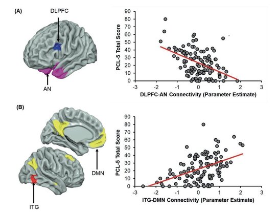
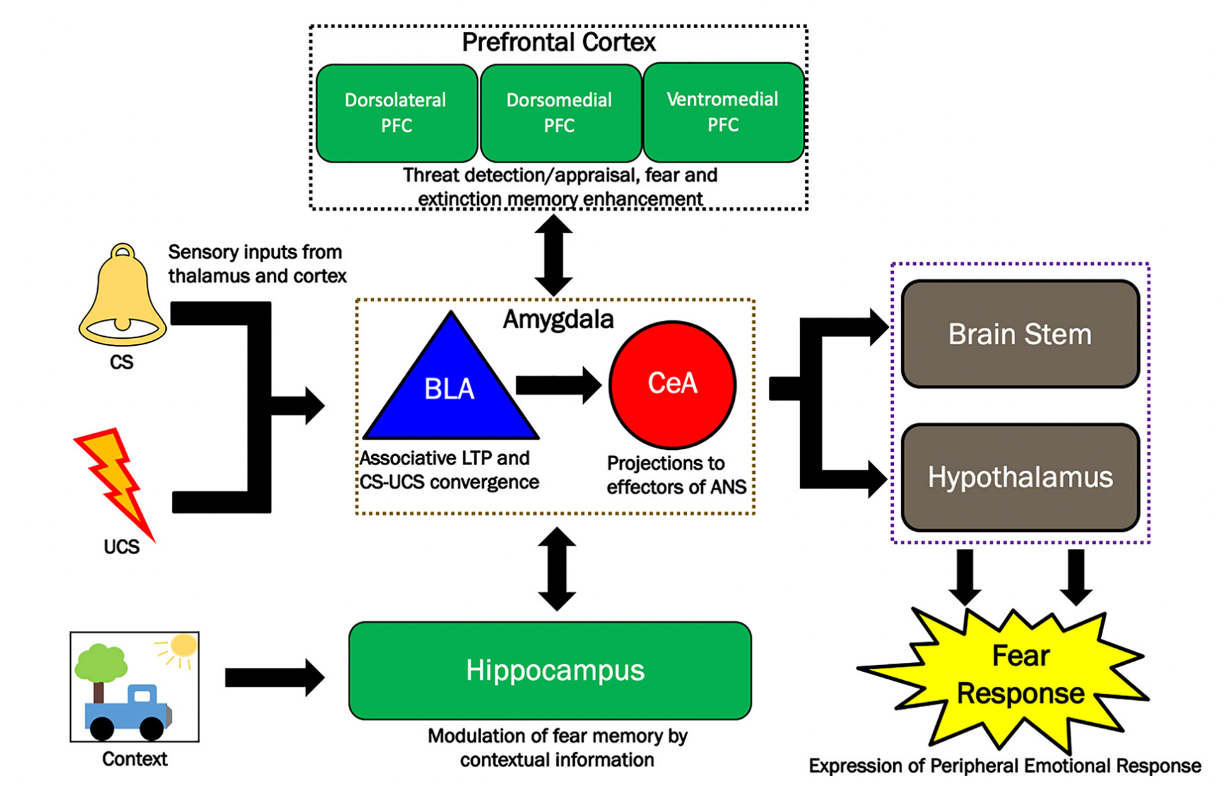
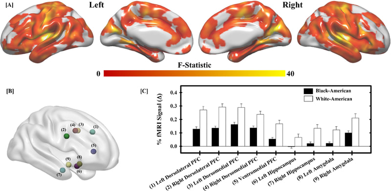
NATHANIEL G. HARNETT, PHD
  • Home
  • Research
  • C.V.
  • Contact

Research

Current Research Direction

​Between 50-90% of people within the United States will experience a traumatic event within their lifetime. Trauma is the antecedent for the development of stress disorders such as Acute Stress Disorder (ASD) and Post-Traumatic Stress Disorder (PTSD). However, not all people who experience a traumatic event go on to develop ASD or PTSD. Specifically, there is considerable individual variability in susceptibility to ASD and PTSD. Individual variability in stress disorder susceptibility may be related to variability in the neurobiology supporting cognitive-affective processes we see disrupted in stress disorder. Therefore, the objective of Nate's research is to understand the neurobiology that mediates susceptibility to trauma and stress-related disorders. Further, the long-term goal is to translate our understanding of the neurobiology of post-traumatic stress into predictive and preventative methods to reduce the incidence of stress disorders.


Approach​

​Nate's research uses a multidimensional approach, incorporating Functional Imaging, Structural Imaging, and Biochemical Assessment to understand the human neurobiology of stress disorder susceptibility. These techniques are combined with psychophysiology and behavioral measures to investigate the relationship between the brain and (dys)functional cognitive-affective processes.


Functional Imaging

​Neural function in Nate's research is assessed using functional magnetic resonance imaging (fMRI) of the blood-oxygen-level-dependent (BOLD) response. BOLD fMRI allows for non-invasive measures of neural activity with humans. Nate's prior work has focused on brain activity underlying emotional learning and memory processes (Harnett et al., 2016; 2015) using Pavlovian fear conditioning procedures. Further, Nate has also investigated how both psychosocial stress (Goodman et al., 2016; Wheelock et al., 2016) and medical trauma (Harnett et al., 2018) alter brain function underlying cognitive and affective behaviors.
Picture
Image adapted from Harnett et al., 2016, Neurobiology of Learning and Memory demonstrating temporally conditioned fMRI signal response within the amygdala.
Structural Imaging

​Nate's research utilizes diffusion-weighted imaging to investigate the white matter architecture connecting brain regions especially important for healthy emotional function (e.g., prefrontal cortex, amygdala, hippocampus, and hypothalamus). Specifically, DWI reconstruction methods allows for visualization and analysis of white matter tracts within the brain. Further, reconstructions of T1-weighted anatomical images are used to assess cortical/subcortical structure properties (e.g., volume, cortical thickness, surface area, etc.). 

Image adapted from Harnett et al., 2018, Brain Imaging and Behavior demonstrating canonical white matter tracts of interest.

Picture
Picture
Biochemical Assessment

​Proton magnetic resonance spectroscopy (1H-MRS) can be used to index concentrations of neurometabolites within the brain such as glutamate/glutamine. Nate's prior work has used 1H-MRS to assess glutamate/glutamine with the dorsal anterior cingulate cortex within traumatized and non-traumatized individuals (Harnett et al., 2017).

Image adapted from Harnett et al., 2017, Journal of Psychiatric Research demonstrating fourier-transformed spectrogram obtained from 1H-MRS.
  • Home
  • Research
  • C.V.
  • Contact